Anatomy of the Eye
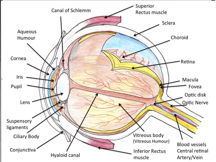
Image adapted from: Virtual Medical Centre.com
The image above is a drawing i have made of the anatomy of the eye
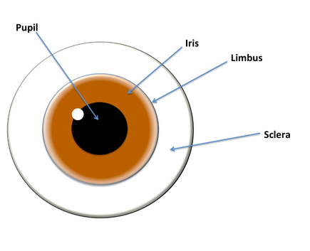
The image above is one i have created, showing the front of the eye
The eyeball consists of three layers:
1. Fibrous layer - This is the external layer and consists of the sclera and cornea. It provides shape and resistance to the eyeball. It also provides an attachment for the extraocular and intraocular muscles of the eye. The sclera covers most of the eyeball whereas the cornea covers a small part of the front of the eye. The sclera is relatively avascular whereas the cornea is completely avascular as it relies on nutrient supply from nearby capillary beds in its periphery, lacrimal fluid and aqueous humour. The junction between the sclera and cornea is called the corneoscleral junction. This comprises the corneal limbus, which is the angle formed by the curvatures intersecting at this region.
2. Vascular layer - This consists of the Choroid, Ciliary body and Iris and is also called the Uveal tract. The choroid is contained between the sclera and the retina and is the vascular part of the eyeball lining most of the sclera. It also contains the choriocapillaries which are closest to, and supply blood to the retina. The choroid is attached to the retinal pigment epithelium. This structure has the highest perfusion rate per gram of tissue compared to all the vascular beds in the body. It is the choroid which causes the 'red eye' to appear in flash photography. The Ciliary body is a muscular thickening which connects the choroid to the iris. It is also an attachment point for the lens. The contraction and relaxation of the ciliary body controls the focussing of the lens by changing its thickness via the suspensory ligaments. Aqueous humour is secreted by the ciliary processes on the internal surface of the ciliary body. This fills the anterior chamber of the eyeball (anterior to lens, ciliary body and suspensory ligaments and behind the iris) and drains into the Canal of Schlemm. The Iris lies anterior to the lens and is the coloured part of the eye. It is composed of two muscles: the Sphincter Pupillae, which constricts the pupil via the parasympathetic nervous system and the Dilator Pupillae, which dilates the pupil via the sympathetic nervous system.
3. Inner layer - This layer is called the Retina and is the sensory neural layer. It consists of two parts: an optic part and a non-visual part.The optic part contains two layers and is sensitive to light. One layer is the neural layer, which is light receptive. The other is a pigmented layer which reduces the scattering of light and therefore enhances the light-absorbing capacity of the choroid layer. The non-visual retina continues anteriorly from the pigmented layer, extending towards the ciliary body and the iris. This is called the ciliary and iridial part of the retina respectively. In clinical terms, the posterior part of the eyeball is called the fundus. An image of a fundus is shown below.
Anatomy of the Retina
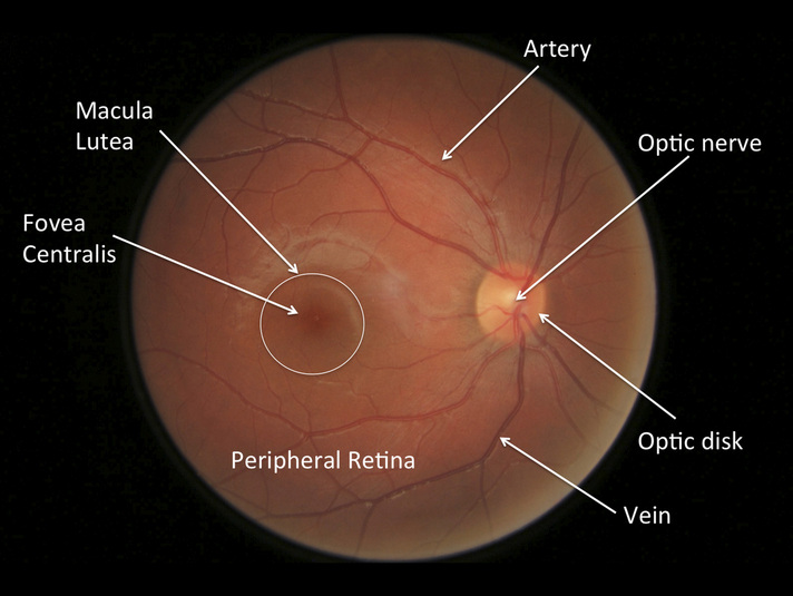
Image adapted from Vision Express Retinal Photography
Above is a labelled image of my own retina, showing the main features of a normal retina
Macula Lutea - This is a rounded yellow area in the retina where the light is focussed on by the lens. This yellow colour is only seen when the retina is being examined by red-free light. It contains the Fovea at the centre and is specialised for the acuity of vision.
Fovea Centralis - This is the a small depression at the centre of the Macula and contains the highest number of photoreceptors, mainly cones, which are involved in high resolution colour vision. This area is avascular and is 1.5mm in diameter. The centre of the fovea is called the foveola.
Optic Disk - This is where the retinal ganglion cells emerge and form the Optic nerve (CN II). The sensory fibers and vessels for the optic nerve enter the eye here. The optic disk contains no photorecepetors and is therefore insensitive to light. This region is called the 'blind spot'.
Peripheral Retina - The peripheral retina contains more rods than cones which are evenly spaced out. This is responsible for peripheral vision.
Blood supply of the Retina -The retina is supplied by the Central Retinal artery which arises from the Ophthalmic artery. The rods and cones on the outer layer are supplied by the choriocapillaries. Venous drainage occurs via the Central retinal vein.
